Optimizing substrates for IR/Raman spectroscopy

Studying cell structures is the foundation of numerous fields in biology including health sciences, pharmacology, microbiology, and more. Traditional microscopic methods of observing structures within cells rely on dyes, fixatives, and/or artificial labels. The full effect that these extra components have on the functions of cell components is still unknown. For example, tacking a green fluorescent protein label to a drug receptor protein may interfere with the receptor binding site and decrease the effectiveness of the drug in the experimental cells (1). Micro-spectroscopy bypasses any interference from labels and dyes and is currently “the only technique for observing molecular activity in humans” (1).
The primary micro-spectroscopic methods currently being applied to cell biology are Raman spectroscopy and Infrared (IR) spectroscopy (2). These techniques rely on vibrational energy in chemical bonds. Raman spectroscopy is more versatile and can be used with a large range of wavelengths and samples in solid, liquid, gas, and gel phases (3, 4). Additionally, IR spectroscopy requires sample preparation whereas Raman spectroscopy can be performed through sample vials or packets (3, 4).
The substrate makes the difference
Visualizing cells with micro-spectroscopy has benefits over traditional microscopic methods. Some of the remaining challenges include achieving a strong signal intensity and reducing background noise. Choosing an appropriate substrate is important to optimize the signal to noise ratio and a spectrum that can be interpreted (5). Different factors that affect substrate choice include the type of spectroscopy, the source wavelength, cost, and reusability (5).
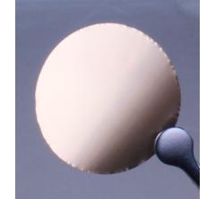
Recommended filter discs for Raman spectroscopy applications are silver membrane filters and gold-coated polycarbonate filters. There is some evidence that silicon, silicon oxide, and highly oriented pyrolytic graphite (HOPG) substrates can yield results in Raman spectroscopy (6), and a study from 2015 reported that Raman-grade calcium fluoride and aluminum-coated substrates gave the most consistent spectra at a range of source wavelengths (5). Coated substrates reduce background noise from uncoated glass and reflective substrates scatter more photons than transparent substrates (5).
IR spectroscopy is compatible with different substrates, depending on the spectral range. Silver and gold-coated membrane filters are recommended for reflectance IR spectroscopy. Aluminum oxide membrane filters work better for transmission IR spectroscopy. Finally, glass microfiber filters (~5 μm) can be used for transmission and reflectance IR spectroscopy.
A future in healthcare
In conclusion, Raman and IR spectroscopy are micro-spectroscopic methods that are being used in new ways to visualize molecules, and choosing an appropriate substrate is a key way to optimize results. Applying these methods to cell biology has significant advantages over traditional microscopic methods because they are label-free, dye-free, and do not require genetic modification of the specimen. Furthermore, molecular analysis of human cells has far-reaching benefits for prediction or early detection of diseases like cancer and Alzheimers as well as ongoing health analysis for patients with chronic conditions (7).
Reference
- Sabbatini S, Conti C, Orilisi G, Giorgini E. Infrared spectroscopy as a new tool for studying single living cells: Is there a niche?. Biomed Spectrosc Imaging 2017 Dec 17; 6(3-4):85-99. https://doi.org/10.3233/BSI-170171
- Matthäus C, Bird B, Miljković M, Chernenko T, Romeo M, Diem M. Infrared and Raman Microscopy in Cell Biology. Methods Cell Biol 2010 Mar 2; 89: 275-308. https://dx.doi.org/10.1016%2FS0091-679X(08)00610-9
- Deepak. Scope of applications of Raman Spectroscopy. Lab-training.com 2015 Jun 30. Retrieved from https://lab-training.com/2015/06/30/scope-of-applications-of-raman-spectroscopy/
- Deepak. Scope of applications of Raman Spectroscopy. Lab-training.com 2015 Jun 30. Retrieved from https://lab-training.com/2015/06/30/scope-of-applications-of-raman-spectroscopy/
- Kerr LT, Byrne HJ, Hennelly BM. Optimal choice of sample substrate and laser wavelength for Raman spectroscopic analysis of biological specimen. Anal. methods 2015 May 15; 7(12):5041-5052. https://doi.org/10.1039/C5AY00327J
- Mikoliunaite L, Rodriguez RD, Sheremet E, Kolchuzhin V, Mehner J, Ramanacicius A, et al. The substrate matters in the Raman spectroscopy analysis of cells. Sci Reports 2015 Aug 27; 5: 13150. https://doi.org/10.1038/srep13150
- Sato H, Ishigaki M, Taketani A, Andriana B. Raman spectroscopy and its use for live cell and tissue analysis. Biomed Spectrosc Imaging 2018 Jan 1; 7(3–4): 97–104. https://doi.org/10.3233/bsi-180184
- Most Viewed Blog Articles (5)
- Company News (285)
- Emerging Technologies (64)
- Microbiology and Life Science News (93)
- Water and Fluid Separation News (97)
- Filtration Resources (93)
- Product News (19)


![Join Sterlitech at BIO 2024 [Booth #5558]: Exploring the Future of Biotechnology](https://www.sterlitech.com/media/blog/cache/300x200/magefan_blog/b4.jpeg)


